Verhoeff-Van Gieson VVG: A Crucial Staining Protocol for Elastic Fibers
The Verhoeff-Van Gieson VVG staining protocol has emerged as a vital tool in the field of histology, particularly for the visualization of elastic fibers in tissue samples. This technique has been widely adopted in research and diagnostic settings due to its ability to selectively stain elastic fibers, providing valuable insights into tissue structure and function. With its applications spanning various fields, including medicine and biology, the Verhoeff-Van Gieson VVG staining protocol has become an essential component of modern histological analysis.
Context and History
The Verhoeff-Van Gieson VVG staining protocol was developed to address the need for a reliable method to visualize elastic fibers in tissue samples. Elastic fibers, which are composed of elastin and fibrillin, play a crucial role in maintaining tissue elasticity and integrity. The development of this staining protocol has enabled researchers to study the distribution and organization of elastic fibers in various tissues, including skin, lungs, and blood vessels. 
Technical Details and Applications
The Verhoeff-Van Gieson VVG staining protocol involves a series of steps, including fixation, staining, and counterstaining. The protocol uses a combination of dyes, including Verhoeff's iodine and Van Gieson's stain, to selectively stain elastic fibers. This technique has been applied in various fields, including cardiovascular research, where it has helped to elucidate the role of elastic fibers in blood vessel function and disease. For example, 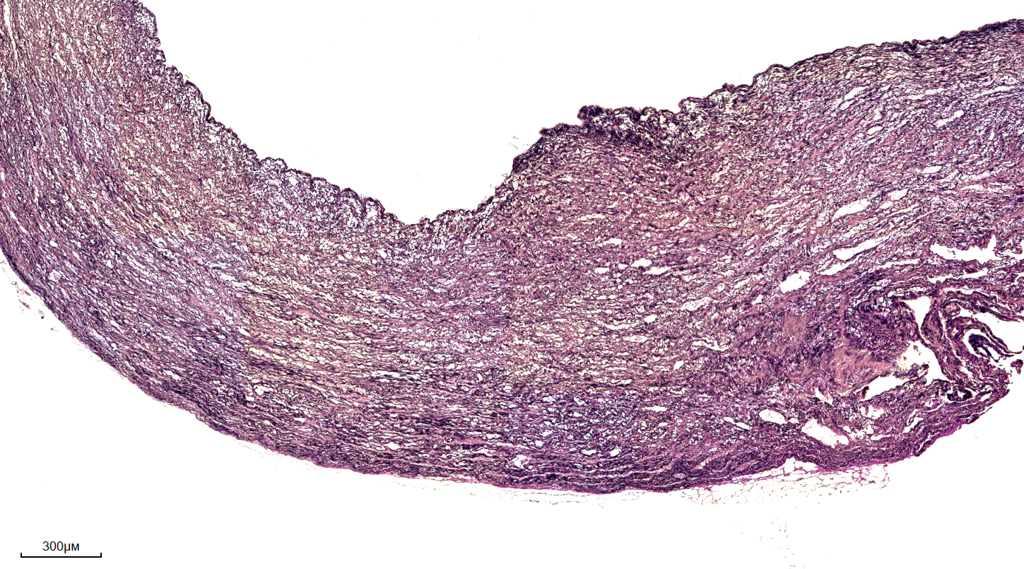
Studies have used the Verhoeff-Van Gieson VVG staining protocol to investigate the distribution of elastic fibers in mouse arteries, providing valuable insights into the relationship between elastic fibers and blood vessel function. The protocol has also been used to study the effects of various diseases, including atherosclerosis and aneurysms, on elastic fiber distribution and organization.
Implications and Future Directions
The Verhoeff-Van Gieson VVG staining protocol has significant implications for our understanding of tissue structure and function. By providing a reliable method for visualizing elastic fibers, this technique has enabled researchers to investigate the role of elastic fibers in various diseases and conditions. As research continues to uncover the importance of elastic fibers in maintaining tissue health, the Verhoeff-Van Gieson VVG staining protocol is likely to remain a crucial tool in the field of histology. Furthermore, the development of new staining protocols and techniques will likely build upon the foundation established by the Verhoeff-Van Gieson VVG staining protocol, enabling researchers to explore new frontiers in tissue biology and disease research.
- The Verhoeff-Van Gieson VVG staining protocol has been widely adopted in research and diagnostic settings due to its ability to selectively stain elastic fibers.
- The protocol has been applied in various fields, including cardiovascular research, where it has helped to elucidate the role of elastic fibers in blood vessel function and disease.
- Future research is likely to focus on the development of new staining protocols and techniques, building upon the foundation established by the Verhoeff-Van Gieson VVG staining protocol.
In conclusion, the Verhoeff-Van Gieson VVG staining protocol has emerged as a vital tool in the field of histology, providing a reliable method for visualizing elastic fibers in tissue samples. As research continues to uncover the importance of elastic fibers in maintaining tissue health, this technique is likely to remain a crucial component of modern histological analysis.
Verhoeff-Van Gieson (VVG) Staining Protocol For Elastic Fibers - IHC WORLD
Verhoeff-Van Gieson (VVG) Staining Protocol for Elastic Fibers - IHC WORLD
Verhoeff Van Gieson For Elastic Fibers – DDK ITALIA

Verhoeff Van Gieson for elastic fibers – DDK ITALIA
Hematoxylin-eosin (HE) And Verhoeff-Van Gieson (VVG) Image After 7
Hematoxylin-eosin (HE) and Verhoeff-Van Gieson (VVG) image after 7 ...